

Saiba como utilizar o AP Zoom
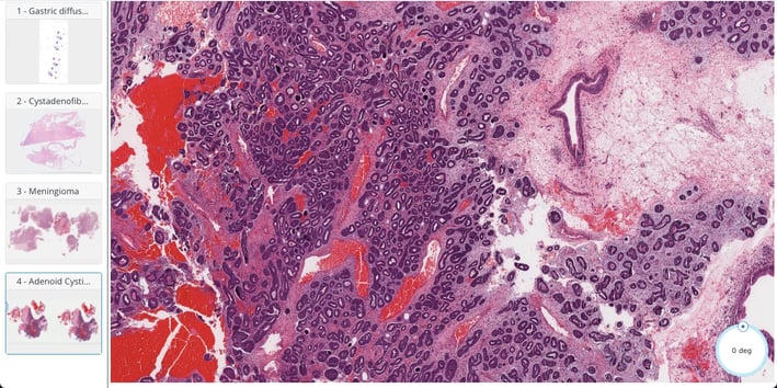
Aprenda a navegar pelas lâminas digitais com facilidade usando o recurso AP Zoom. Amplie, explore detalhes morfológicos e visualize as anotações interativas em alta resolução. Uma ferramenta essencial para o estudo dinâmico e preciso da histopatologia.
Sobre Nós
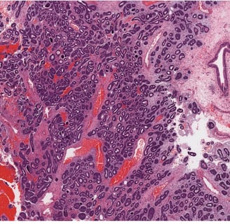
O AP Zoom é uma iniciativa educacional desenvolvida com o objetivo de aprimorar o ensino e a aprendizagem na área da patologia por meio de recursos digitais interativos. Este projeto reúne uma curadoria de lâminas histopatológicas de casos reais, cuidadosamente selecionadas por sua relevância diagnóstica e valor pedagógico.


Atlas Digital
Desenvolvimento de um atlas digital para ensino em patologia com lâminas histopatológicas interativas.


Contato
Entre em contato para mais informações sobre nosso atlas digital.