Atlas Digital

Um atlas interativo para ensino em patologia e histopatologia.

A tablet displaying an educational diagram of a human eyeball resting on a dark sofa with a light cushion behind it. A blue-green textured blanket is draped around the tablet, and a white stylus is placed beside it.
Lâminas Histológicas

Cada lâmina digital inclui anotações e perguntas para autoavaliação, promovendo um aprendizado ativo e acessível para estudantes de pós-graduação em patologia e áreas relacionadas.

The image depicts a microscopic view of a cellular structure with numerous circular blue elements scattered throughout a textured, grayscale background. The scene resembles a cross-section of complex organic material with various bubble-like formations.
Estudo Ativo

O projeto visa facilitar o ensino de patologias com um ambiente visual e interativo, organizando casos relevantes por categorias morfológicas e sistemas, enriquecendo a experiência educacional dos alunos.

Two people in lab coats collaborate in an office setting. One is seated and pointing at a computer screen displaying medical images, while the other stands nearby holding a tablet. Large windows reveal a view of greenery outside, creating a bright and professional environment.

Atlas

Explore casos histopatológicos interativos para aprendizado em patologia.

A detailed anatomical model of the human foot showing bones, muscles, tendons, and ligaments. The model is sectioned to reveal the internal structure and is labeled with numbers for educational purposes. The surface texture imitates human tissue, highlighted with colored sections to differentiate between various anatomical features.
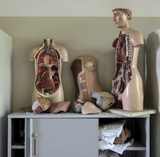
Two anatomical models displaying the human torso are positioned on a cabinet. Cabinets on either side hold various medical supplies and material, with some shelves visible through glass doors. Loose anatomical parts, including a heart and sections of the torso, are scattered around.
An illuminated anatomical representation with vibrant red and pink tones, resembling a stylized, artistic depiction of a skull or skull-like structure with an intricate pattern. The symmetrical design evokes a sense of mystery and scientific exploration.
A close-up view of cells under a fluorescent microscope, showing bright green and blue colors. The cells have distinct blue nuclei surrounded by an irregular green cytoplasm, all set against a dark background.
A detailed anatomical model of a human heart featuring prominent red and blue blood vessels. The model shows the heart's intricate structure and various components in a realistic and educational depiction.